Wednesday 7 December 2011

Neuropsychological Assessment

What is it ????
NeuropsychologicaI Assessment is an appraisal of the brain's psychological, that is cognitive and behavioural, functions.Neuropsychological assessments are used to assess the level of impairment to particular skills and locate the area of the brain affected by damage after brain injury or a neurological illness. Neuropsychological assessment is the administration of neuropsychological tests to assess cognitive functioning. Aspects of cognitive function that are usually assessed include, intelligence, language, visuo-perception, memory and executive
So how are they done???

Methods of assessment can include Interviews and behavioural observation,Normal psychometric tests e.g. IQ scales, Standardised neuropsychological tests (e.g. Recognition Memory Tests (Warrington, 1986) )and Unstandardised clinical tests. (e.g. Drawing objects from memory in cases of
unilateral neglect.)
How long does neuropsychological assessment take?
Neuropsychological assessments can vary in time On average it can take between 2-4 hours dependent on what the purpose of he assessment is. It can also vary dependent on the patient’s response time and how they cope with the test and also their age.
So what are the Goals of neuropsychological assessment ????

Contributing to Diagnosis
• Major brain damage is now diagnosed by neuroimaging.
• But diffuse or early cortical damage may only be evident
from cognitive dysfunction (e.g. AD, head injury).
Guiding management or rehabilitation.
• Impossible to predict functional problems directly from
brain scans.
• Neuropsychological assessment is crucial in identifying
deficits and spared functions.
Monitoring recovery or deterioration.
• A significant degree of recovery occurs over the months
after non-degenerative damage (head injury, stroke).
•Deterioration in degenerative conditions may be slowed by
medical treatments (e.g. drugs in AD).
•Need for serial neuropsychological assessments.

Friday 2 December 2011

Memory !!

Memory is the process used to acquire store and retain information that can later be retrieved. There are three major processes involved in memory. ENCODING, STORAGE AND RETRIEVAL. Memory cannot be found in one specific area of the brain as it a process nit a unitary entity.However the hippocampus is involved in the consolidation of a memory  and damage to this area can cause impairments between STM and LTM. 
Encoding is the 1st step in the creation of a memory and is rooted in the senses. Each separate sensation of an event travels to the hippocampus these perceptions of the event are then integrated and stored into one single experience.
There are many subtypes of memory illustrated in the picture below.
Storage of memories retention of information, which has been achieved through the encoding process, in brain for prolonged period of time until it is accessed by the recall process. Mmemory can be stored in STM or LTM (read further info for more on this) 
Retrieval of memories is the process of accessing stored memories. Once information has been encoded and stored in memory. It must be retrieved in order to be used.

The causes of amnesia can be divided into categories. Memory appears to be stored in several parts of the limbic system of the brain, and any condition that interferes with the function of this system can cause amnesia. The types of amnesia are
§  Anterograde amnesia, is the loss of short-term memory, the loss or impairment of the ability to form new memories through memorization. People may find themselves constantly forgetting a piece of information, people or events after a few seconds or minutes, because the data does not transfer successfully from their conscious short-term memory into permanent long-term memory. Primarily in older men, transient global amnesia causes severe loss of memory for minutes or hours.
§  Retrograde amnesia, the loss of pre-existing memories to conscious recollection, beyond an ordinary degree of forgetfulness. The person may be able to memorize new things that occur after the onset of amnesia (unlike in anterograde amnesia), but is unable to recall some or all of their life or identity prior to the onset. The affects of retrograde amnesia (RA) occurs on fact memory on a lower degree than its affects on autobiographical memory, which can be affected over the whole lifespan of the patient by RA

Further info: 
Sensory Memory Sensory memory is the earliest stage of memory. During this stage, sensory information from the environment is stored for a very brief period of time, generally for no longer than a half-second for visual information and 3 or 4 seconds for auditory information. We attend to only certain aspects of this sensory memory, allowing some of this information to pass into the next stage - short-term memory.

Short-Term Memory Short-term memory, also known as active memory, is the information we are currently aware of or thinking about. In Freudian psychology, this memory would be referred to as the conscious mind. Paying attention to sensory memories generates the information in short-term memory. Most of the information stored in active memory will be kept for approximately 20 to 30 seconds. While many of our short-term memories are quickly forgotten, attending to this information allows it to continue on the next stage - long-term memory.

Long-Term Memory Long-term memory refers to the continuing storage of information. In Freudian psychology, long-term memory would be call the preconscious and unconscious. This information is largely outside of our awareness, but can be called into working memory to be used when needed. Some of this information is fairly easy to recall, while other memories are much more difficult to access.


Tuesday 22 November 2011

Frontal Lobes !


One of four main regions of the cerebral cortex is the frontal lobes. They are at the front most region of the cerebral cortex. The frontal lobes are involved in problem solving, decision making, movement and planning. The frontal lobe has 3 main division consisting of the prefrontal cortex and the pre-motor area and the motor area. The pre-motor and motor areas of the frontal lobes contain nerves that control execution of voluntary muscle movement and the prefrontal cortex is responsible for personality and expression and the planning of complex cognitive behaviour..
Although the frontal lobe is the largest lobe in the brain however in routine neurologic examinations it is not usually specifically evaluated as cognitive tasks usually require multiple brain regions both within and outside the frontal lobes and so therefore it can be difficult during examinations. Dysfunctions or damage to the frontal lobes can cause relatively specific clinical syndromes. In most cases when a patients history suggests that they have a dysfunction of the frontal loves neurobehavioral evaluation is necessary.
Lesions of the frontal lobes and deeper brain structures generate relatively distinctive clinical behaviors such as these:


  • The dorsolateral frontal cortex is concerned with planning, strategy formation, and executive function. Patients with dorsolateral frontal lesions tend to have apathy, personality changes, abulia, and lack of ability to plan or to sequence actions or tasks. These patients have poor working memory for verbal information (if the left hemisphere is predominantly affected) or spatial information (if the right hemisphere bears the lesion brunt).
  • The frontal operculum contains the center for expression of language. Patients with left frontal operculum lesions may demonstrate Broca aphasia and defective verb retrieval, whereas patients with exclusively right opercular lesions tend to develop expressive aprosodia.
  • The orbitofrontal cortex is concerned with response inhibition. Patients with orbitofrontal lesions tend to have difficulty with disinhibition, emotional lability, and memory disorders. Patients with such acquired sociopathy, or pseudopsychopathic disorder, are said to have an orbital personality. Personality changes from orbital damage include impulsiveness, puerility, a jocular attitude, sexual disinhibition, and complete lack of concern for others.
  • Patients with superior mesial lesions affecting the cingulate cortex typically develop akinetic mutism.
  • Patients with inferior mesial (basal forebrain) lesions tend to manifest anterograde and retrograde amnesia and confabulation.
The image below shows an MRI that is suggestive of frontotemporal dementia:

Friday 11 November 2011

Main things to remember about emotion!!

Emotion is a literal Latin translation that means “to move” or “to stir up” Emotion is different from “mood” and “affect”. Brewin (1988) is a broad term and subsumes much behaviour, including mood, feeling, attitude, preferences and evaluations.


Mood= Refers to the frame of mind or “emotional state” of a person which is defined by the individuals internal state and not external behaviour.

Emotions= Thought to be briefer more spontaneous and detectable from the appearance of the organism.
The subcortical structures are thought to be involved in regulation of emotion is the hippocampal system and the amygdale.
The cognitive appraisal theory states that emotions are judgements about the extent that the current situation meets your goals. William James argued that emotions are perceptions of changes in the body like heart rate, breathing rate, perspiration and hormone levels. Happiness is a physiological perception, not a judgement and other emotions like anger and sadness are mental reactions to different kinds of physiological stages.
  Measuring and maniuplating emotions
Tools used to measure emotion is the accelerometers for motion, skin conductance sensors to measure excitement level, temperature sensors and also facial recognition through the use of simple webcams.
Emotion is also assessed and manipulated through mood induction by using thought ( getting the person to think of something) to achieve a required state, through the use of films and music a particular mood can be achieved. Reward and punishment using reinforcer’s to trigger an emotion. Emotions can also be manipulated by loud noises, mild electric shocks, words that represent emotional concepts or pictures of emotional scenes.
Examle of emotion testing Lowa Gambling Task
Lowa Gambling Task was created to stimulate real life decision making. The task was introduced Antonion Damasio. The task was presented as a gambling task it looks at the cognition and emotion.The task consists of participants are presented with 4 virtual decks of cards on a computer screen. They are told each time they choose a card they will win some money in the game. Once in a while choosing a card causes the participant to lose some of the money they have earned in the game. The main goal of the game is to gain as much money as possible to win. Every card drawn will earn the participant a reward and sometimes a card could cause them a penalty. Some decks are “bad decks” and some are “good decks” some will lead to more losses than wins over the period of the game and others will lead to more gains and wins. The decks differ from each other in the number of trials.

Wednesday 26 October 2011

Movement Disorders


Movement Disorders
1ST A bit of info on the framework  of movement !
The motor cortex
The motor cortex is one area of the brain which is most involved with controlling voluntary movements. The motor cortex is located in the back portion of the frontal lobe.The motor cortex is divided into two main areas. the primary motor cortex and the somatiosensory cortex.  For voluntary movement to be carried out the motor cortex must 1st receive information about the bodys position in space from the parietal lobe about the movement that needs to achieved and the appropriate method for attaining it, from the anterior portion of the frontal lobe about memories of previous strategies.
Basal Ganglia
The basal ganglia is found deep inside the cerebum the main areas of the basal ganglia are the caudate nucleus, the putamen, and the globus pallidus.These are clusters of nerve cella which are interconnnected tightly they receive information from several different regions of the cerebral cortex. Once the basal ganglia collects and processes this info the send it to the motor cortex through the thalamus.
This diagram below shows the system of communication in the brain
In addition, to ensure that all of these movements are fast, precise, and co-ordinated, the nervous system must constantly receive sensory information from the outside world and use this information to adjust and correct the hand's trajectory. The nervous system achieves these adjustments chiefly by means of the cerebellum, which receives information about the positions in space of the joints and the body from the proprioceptors.

Parkinsons disease
Parkinsons disease affects the way in which the brain co-ordinates body movements inlcuding walking talking and writing.
The cause of parkinsons disease is a chronic degenrative disorder of the parts of the brain that control motor system and manifests with progressive loss of the abiklity to co ordinate movements. This happens when  the loss of nerve cells In the area of the brain called the substantia Nigra of the mid brain area (controls movement) dies or suffers from some damage. Because of these dying cells the chemical messenger dopamine is no longer sufficent in the amount that is produced and so the onset of  sympotms such as a resting tremor, slowness in initiating movement and muscle stiffness can occur. This causes movement does not work so well and causes it to become abnormal or slow.
The process of the loss of the nerve cells is a slow provess and the level of dopamine in the brain will fall over a period of time 80% of the nerve cells in the substantia nigra have gone then the symptoms begin to appear and gradually begin to worsen
The diagram above shows exactly where the substantia nigra is found in the brain.


Tourettes syndrome
Although the cause of TS is unknown, current research points to abnormalities in certain brain regions (including the basal ganglia, frontal lobes, and cortex), the circuits that interconnect these regions, and the neurotransmitters (dopamine, serotonin, and norepinephrine) responsible for communication among nerve cells. Given the often complex presentation of TS, the cause of the disorder is likely to be equally complex.


Symptoms

Tics are classified as either simple or complex. Simple motor tics are sudden, brief, repetitive movements that involve a limited number of muscle groups. Some of the more common simple tics include eye blinking and other vision irregularities, facial grimacing, shoulder shrugging, and head or shoulder jerking.  Simple vocalizations might include repetitive throat-clearing, sniffing, or grunting sounds. Other complex motor tics may actually appear purposeful, including sniffing or touching objects, hopping, jumping, bending, or twisting. Simple vocal tics may include throat-clearing, sniffing/snorting, grunting, or barking. More complex vocal tics include words or phrases.  Perhaps the most dramatic and disabling tics include motor movements that result in self-harm such as punching oneself in the face or vocal tics including coprolalia (uttering swear words) or echolalia (repeating the words or phrases of others). Some tics are preceded by an urge or sensation in the affected muscle group, commonly called a premonitory urge. Some with TS will describe a need to complete a tic in a certain way or a certain number of times in order to relieve the urge or decrease the sensation.
Tics are often worse with excitement or anxiety and better during calm, focused activities. Certain physical experiences can trigger or worsen tics, for example tight collars may trigger neck tics, or hearing another person sniff or throat-clear may trigger similar sounds. Tics do not go away during sleep but are often significantly diminished.


Wednesday 19 October 2011

Hemispatial Neglect and Blindsight

Hemispatial neglect – This is the failure to be aware of objects to one side of space. This is usually the contralesional side so if the righ side of the brain has been affected by a lesion than the neglect will be to the left and if the left side of the brain has been affected by a lesion the neglect will be to the right.

Neglect is most prominent and long lastinig in right hemisphere lesions of the brain and this can be especially after a stroke.

(Below is the link to a video of an example of a person whp has hemi spatial neglect drawing a picture)


Cognitive deficits underlying neglect

è Many different cognitive deficits either alone or combined

è Deficit in directing attention to the left

è Impaired representation of space

è Direcetional motor impairment patients could experience difficulty in initiating or programming leftward movements In addition to these lateralised impairments (worse tothe left following right-hemisphere stroke), neglect syndrome also consists of non-spatially lateralised deficits, involving both sides of space. Different patients may suffer different combinations of lateralised and non-lateralised deficits, depending upon the precise location and extent of their lesions.

è Impairments in sustained attention

è Bias to local features in the visual scene

è Deficit in spatial working memory

è Prolonged time- course of visual processing


Blindsight is the term used when people are perceptually blind in particular area of the visual field they are able to respond to visual stimuli. Blindsight is caused by injury to the occipital lobe this is the part of the brain that is responsible for vision. Type 1 blindisght subjects have no awareness to any stimuli but there able to make prediction which are at levels to high to be by chance aspects of visual stimulus such as location or the type of movement a stimulus is displaying this is usually in situations where response is forced or in a situation where they must guess. Type 2 blindsight is when the patient has awareness of movement within the blind area but they have no visual perception. This could be due to the patient being aware of their eyes tracking motion which all the person is blindsighted will still function normally.

(An example and explanation further of blindsight can be seen in the video below) http://www.youtube.com/watch?v=sq6u4XVrr58&list=PL361F982E5B7C1550&index=1

Thursday 13 October 2011

Visual Perception Disorders

Agnosia = loss of knowledge. It is the loss of the ability to recognize objects, persons, sounds, shapes, or smells while the specific sense is not defective or is there any significant memory loss. Bauer (1993) defined agnosia as a “failure of recognition that cannot be attributed to elementary sensory defects mental deterioration, attentional disturbances, aphasic misnaming or unfamiliarity with sensorially presented stimuli”

Brain damage can affect a variety of sensation such as determining the presence of absence of light, detecting changes in contrast, discriminating between or perceiving colour. Diagnosis of agnosia dictates that these sensory deficits are absent.

There are a variety of different types of agnosia Lissauer (1890), agnosia manifested itself in two distinct forms: appreciative agnosia & associative agnosia. Two types of agnosias are visual agnosia  and object agnosia. Visual agnosia is the severe inability to recognize visual stimuli despite sensory abilities. Object agnosia is the inability to identify objects that are presented, in these case they can neither name or give other evidence of recognizing visually presented objects. Prosopagnosia is another form of agnosia and is sometimes also called faceblindness and facial agnosia. Patients cannot consciously recognize familiar faces, and can at times even be their own. It can sometimes be perecievd as an inability to remember names.


http://www.youtube.com/watch?v=rwQpaHQ0hYw  < - Object agnosia

http://www.youtube.com/watch?v=OYW8vJ5232o <- Visual agnosia

http://www.youtube.com/watch?v=ZogbIvdgfzQ&feature=related <- Prosopagnosia

Wednesday 12 October 2011

Tools of the trade ! Human neuropsychology

Over many years neurologists and psychologists have been interested in exactly what it is that causes people to behave the way they do in everyday behaviour and people with mental illnesses or brain damaged patients. With the use of a variety of different tools to identify specifically what areas of the brain  is involved in different actions. 
It is most common for neurologists or psychologists to assess damaged brains as it allows clinicians to contrast the impaired abilities to the intact ones, this also helps when it comes to rehabilitation as clinicians can target the intact abilities to help the patient. 
Past brain assessments such as MR scans reveals that the brain is completely normal and Lesions do not show everything. PET and FMRI and NMR shows areas of activity and inactivity allowing you to assess behaviour and link to highlighted areas of the brain.
Hysterically paralysed patients where asked to attempt to move paralysed area of body even if the body can’t  the PET scan then highlights in brain the active areas. Through the use of EEG  researchers are able to test patients reaction time. Researchers could tell the patient to move their finger and the EEG shows that before the finger is moved the area required to move the finger is highloighted and there is a second delay before the action is executed, it has been suggested that this delay is dued to neural processing.
There are six different types of tools that are used to look at the process
Single Case Studies
This is the investigatiom of behaviour of one individual over a intensively long period of time.


Advantages
è Valuable because human brain lesions cannot be performed experimentally
è Brain damage may highlight role of damaged region in function
è Allows in-depth study over long periods of time
è Can be used to constrain theproespf cognition in a way not possible in experimental studies of healthy individuals
Disadvantages
è Invasive
è Subject to individual differences
è Locus of damage can be variable and may not always be described accurately
è Previous level of functioning may be unkown
è Other damaged brain regions may be producing deficits
è There may be confounding factors such as medication use
Electroencephalography (EEG)
This tool records electroencephalograms also known as brainwaves. The method is non invasive as brainwaves are gathered through the use of electrodes which are placed on the scalp.


Advantages
è Non-invasive
è Excellent temporal resolution
è Relatively easy and cheap to operate
è Can be used with healthy and clinical human participants
è Can be used to record brain electrical activity in real time
è Can be used to measure the brains response to a number of psychological variables
Disadvantages
è Ability to localize function is weak – generating source may be at some distance from the recording source
è Activity is recorded from millions of groups of neurons
è Signal may be attenuated and smeared
è Brain activity may fluctuate unpredictably and ‘chaotically’
è Susceptible to movement artefact
è Unclear what EEG changes signify
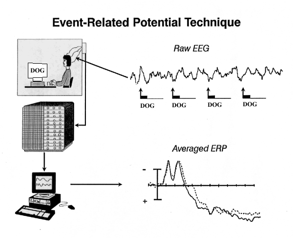
Event Related Potentials (ERP)
Event-related potentials (ERPs) are large slow brainwaves that appear as a result of sensory or cognitive stimulation.


Advantages
è Can be used in healthy and clinical participants
è Useful index of sensory function
è Possible measure of cognition decline and normal cognitive function
è Non-Invasive
è High temporal resolution (one millisecond)
è Relatively easy to use and measure
Disadvantages
è Significance of some waves unknown
è Mechanism underlying ERP poorly understood
è Spatial resolution relatively poor

Positron Emission Tomography (PET)
It measures brain function via the measurement of brain oxygen consumptions blood flow and glucose metabolism. Blood flow is the most reliable of those measurements


Advantages
è Can be used with most clinical and healthy subjects
è High spatial resolution
è Measure of neuronal activity (indexed by blood flow/metabolism) in vivo
è Give three- dimensional representation of regional activity
è Can be used to measure brain activity during task performance
Disadvantage
è Invasive
è Poor temporal resolution (Blood flow is slower than neural transmission)
è Cannot be used in children or premenopausal women (because of the injection of radioactive substances)
è Tasks must take longer than a minute
è Averaging does not take into account contro-anatomical variation
è Expensive

Magnetic Resonance Imagining (MRI)
Magnetic resonance imaging (MRI) is based on changes in the magnetic properties of atoms and was developed to observe the activity of atomic nuclei.


Advantages
è Non-invasive and non- toxic
è Allows structural imaging
è Provides the best spatial resolution of current imaging technique (1-2mm)
è No known biological risk
Disadvantages
è Procedure difficult if participant is claustrophobic
è Magnet precludes introduction of ferromagnetic material into testing environment
è Equipment producing radio frequencies must be shielded
è Obtaining good images from areas near to large cavities is diffuclt
è Transient scanner effects can produce one bad image out of ten or twenty
è As with PET, ensuring similar heard placement for each participant is difficult
è Noisy procedure

Functional Magnetic Resonance Imaging (FMRI)
FMRI measures blood oxygen level dependent ( BOLD) responses . It has been used to study a variety of behaviour, from language difficulties in developmental dyslexia to face recognition, to personaility and the recognition of the emotions to being in love.

Advantages
è Measures direct changes in brain tissue from normal and clinical participants
è Non invasive and non- toxic
è Allows functional imaging
è Provides the best spatial resolution of current imaging techniques (1-2mm)
è No known biological risk
è More widely available and cheaper than PET
Disadvantages
è Decrease in venous oxygen content are not observed by FMRI
è Procedure difficult if participant is claustrophobic
è Poor temporal resolution ( four images per second is the current norm)
è Magnet precludes introduction of ferromagnetic materials into testing environment
è Equipment producing radio frequencies must be shielded